Multi-prior intelligent microscopy assisted quantitative phase imaging
Recently, a research group led by Prof. YAO Baoli and BAI Chen form Xi'an Institute of Optics and Precision Mechanics of the Chinese Academy of Sciences (CAS), developed a training-free Multi-Prior Physics-enhanced Neural Network (MPPN-PSR) for high-throughput, pixel super-resolution quantitative phase imaging. The study was published in Opto-Electronic Advances.
Digital holographic microscopy is commonly used for non-destructive phase imaging and revealing key features of samples. Among these, digital in-line holographic microscopy (DIHM) has relatively higher space-bandwidth product. Unfortunately, twin-images generated during holographic reconstruction process need to be eliminated either by complicated setup or algorithms. In addition, a larger pixel size sensor has high photosensitivity, large field-of-view (FOV), and improved signal-to-noise ratio, but will leads to inadequate sampling of image, resulting in low pixel resolution.
Deep learning (DL) network, with its noise suppression and inverse problem-solving capabilities, has become a powerful tool for DIHM imaging and Pixel Super-Resolution (PSR). However, most DL-based strategies are data-driven or end-to-end net approaches, suffering from excessive data dependency and limited generalization ability.
In this study, the researchers reported a training-free network for quantitative phase retrieval in DIHM imaging with PSR, i.e., the MPPN-PSR, which encapsulates the physical model prior, sparsity prior and deep image prior in an untrained deep neural network. Without any additional hardware design or data training, pixel super-resolution, suppression of both noise and twin-image of phase reconstruction can be achieved from a single-shot DIHM hologram.
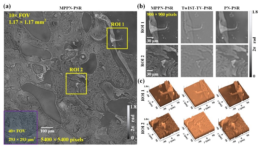
MPPN-PSR maximizes the data efficiency by reducing the intensity image redundancy requirement to only one frame, and improves the space-bandwidth product (SBP) since the inherent large FOV of the low-resolution intensity image is exploited. The imaging FOV is about 1.4 mm2 with increased optical resolution to 1.1 μm while the effective pixel size is improved to 2.2 μm from 6.5 μm.
“These superior performances can be widely used in biomedical workflow and industrial measurement.” said BAI Chen, an expert of intelligent microscopy, and lead author of the study.
(Available Online: 28 August 2024)
Comparison of TOMM20 antibody imaging under different methods. (Image by XIOPM)
